Fully Endoscopic Pineal Tumor Removal at the Skull Base Institute
Pineal tumors are rare and very serious. Complications may include headaches, nausea, fatigue, visual impairments, memory problems and seizures. For this reason, Skull Base Institute’s founder, Dr. Shahinian developed the revolutionary and fully endoscopic “keyhole” procedure to treat pineal tumors. The Skull Base Institute has successfully treated over 6,000 patients.
Feel free to call or Contact Us at any time for answers to your questions or to schedule a treatment consultation.
To help our visitors understand this difficult condition, we encourage you to continue reading about pineal tumors and treatment below. The colorful buttons above will also guide you to a procedural video, published articles and dozens of patient testimonials. You can even see Dr. Shahinian on Ellen, NBC and The Doctors Show at our Videos & Animations page! The entire staff at Skull Base Institute is passionate about treating pineal tumors so please contact us so we can help you.
Because of the pineal gland’s location deep in the midbrain area, traditional open surgery is often times a very aggressive and intrusive procedure. Even the process of accessing the tumor stereotactically to perform a biopsy can be quite involved, subjecting the patient to the risk of hemorrhage or direct neurologic injury. Instead of open brain surgery or stereotactic biopsy, the Skull Base Institute has pioneered a different method: using tiny endoscopes to reach the tumor directly and excise it completely. Patients receive all the advantages of this revolutionary breakthrough in the field of brain surgery, such as no retraction or disruption of brain tissue, faster recovery time, less pain, less scarring and significantly reduced chance of complications.
The goal is that the brain remains virtually untouched, except for the tumor that’s been removed. As Dr Shahinian says himself, “You want to go in and out without the brain knowing you were there.”
Overview
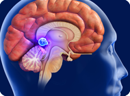
The pineal gland is a small gland in the mid-brain shaped like a pine cone. It produces melatonin, a serotonin derivative and hormone that controls sleep/waking patterns, circadian rhythms and the body’s ability to regulate to the seasons.
Pineal tumors are rare in adults, representing 1% of all brain tumors overall. However, pineal tumors make up 3-8% of intracranial tumors in children and the average age of patients at time of diagnosis is 13 years old.
The most common types of pineal tumors are germ cell tumors (germinoma, teratoma); glial cell tumors (astrocytoma, ependymoma); pineal cell tumors (pineocytoma, pineoblastoma) and miscellaneous tumors (pineal cyst, meningioma). 10% of pineal tumors are benign, another 10% are relatively benign (low grade), while the remaining 80% are highly malignant.
In children younger than 10, certain germ cell tumors may secrete hormones that can create an endocrinologic disturbance and lead to precocious puberty. In this instance, the pineal tumor affects the hypothalamus, which in turn can compromise or overstimulate pituitary function.
Causes
As with most brain tumors, the exact causes of pineal tumors are frequently unknown. Most pineal tumors arise sporadically and are not genetically linked. The events that begin or trigger the abnormal development are not fully understood.
Typically, the pineal tumor starts out small, (and asymptomatic), but can grow at varying rates over time. They are more common in children.
Symptoms
In general, symptoms will vary in severity depending on several factors: the type and size of the pineal tumor, whether the tumor interferes with hormone levels or the flow of the cerebrospinal fluid (CSF), and whether the tumor is causing pressure on surrounding vital structures.
Symptoms in children and adults may include headaches, nausea, fatigue, visual impairments, double vision, memory problems, and seizures. In children, precocious puberty has been identified as a key indicator of a pineal tumor.
In some cases, although the tumor itself may not be malignant, its location deep in the brain represents a problem. The pineal tumor can press on neighboring neurovascular structures and, due to its proximity to the aqueduct of Sylvius, may lead to hydrocephalus or increased intracranial pressure.
Diagnosis
In addition to a full medical history and physical examination, diagnostic tools can include the following:
Magnetic resonance imaging (MRI) is the most important diagnostic tool for tracking the size, shape and precise location of the pineal tumor, as well as its tendency for growth.
A computed tomography (CT) scan can identify the degree of hydrocephalus (pressure that builds up within the cerebrospinal fluid) and whether the tumor is calcified.
MRA (Magnetic Resonance Angiography) uses contrast material to provide a look at blood vessels in the body by mapping blood flow and the condition of the blood vessel walls. In many cases, MRA can yield information that adds to the information gained by an MRI and therefore these two studies (MRI/MRA) are done in conjunction.
For some pineal tumors (germinomas), analysis of the cerebrospinal fluid (CSF) can reveal high levels of certain markers (Beta-HCG, AFP and CEA).
Treatment
Because of the remote and difficult-to-reach location of the pineal gland, observation (the “wait and see” approach) is often advised. When treatment is necessary, the traditional approaches involve either one of the open brain approaches or a stereotactic biopsy combined with either radiotherapy, chemotherapy or both.
Although microsurgical advancements have led to better outcomes related to this type of surgery, due to the deep location of pineal tumors, it is considered one of the most difficult types of open brain surgery.
In contrast, at the Skull Base Institute, we advocate the minimally invasive endoscopic approach as the mainstay of therapy. This curative procedure begins by creating a “keyhole” smaller than a dime behind the ear. From there, a slender endoscope slides over the top of the cerebellum and through a natural pathway to access the deep-seated pineal tumor, without the need for any metal retractors or the need to go through normal brain tissue. This revolutionary procedure is used to completely excise the tumor.
Prognosis
Pineal tumors are treatable, and in many cases, curable. The ideal approach to treatment depends on the size and type of tumor, and the extent to which the tumor is invading or compressing surrounding neurovascular structures.
Where surgical intervention is indicated, the minimally invasive endoscopic approach leads to a shorter surgery and recovery times, less pain and fewer complications than traditional types of open brain surgery.
If you or someone you love has questions about Pineal Tumors, please Contact Us and we will be happy to help you.
