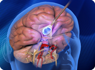
Meningioma: Minimally Invasive, Tailored Meningioma Surgery
Tumors once thought to be “unresectable” are now regularly and safely removed at the Skull Base Institute. Meningiomas located at the base of the skull are very difficult to access. Our use of highly specialized surgical techniques, sophisticated intraoperative monitoring equipment, and minimally invasive surgical instruments allow us to expose hard-to-reach areas in their entirety without disturbing surrounding critical neurovascular structures.
Overview
Meningiomas are tumors that originate from the meninges which are membrane-like structures that surround the brain and spinal cord. The dura, which is the outermost (and toughest) of the meninges covers the entire interior of the skull and invaginates into the brain creating “dural folds” and compartments within the interior of the skull such as the falx cerebri which separates the right side of the brain from the left side and the tentorium cerebelli which forms a “tent” that separates the cerebrum from the infratentorial space that contains the cerebellum and the brainstem within the posterior cranial fossa.
Typically, meningiomas are benign and slow-growing tumors, but instances of malignancy have been reported. Only about 10% of meningiomas occur in the spinal cord, while the majority occur intracranially. Intracranial meningiomas may occur in variable locations of the brain as dural-based, dome-shaped, round or oval, solitary masses. Rarely multiple concurrent meningiomas occur simultaneously in different locations of the brain. An unusual appearance for meningioma, called meningioma-en-plaque, has a flattened appearance that conforms to the curves of the brain and the inside of the skull.
Meningiomas represent the second most common type of tumor of the brain, accounting for approximately 20% of all primary intracranial tumors in adults. They are most common between the ages of 40 and 70 and they are more common in women than in men. Among middle-aged patients, there is a marked female predominance, with a female:male ratio of approximately 3:1. Meningiomas are extremely rare in children and only 1.5% of the total cases of meningiomas occur in the pediatric population.
Causes
The only known predisposing factors associated with the occurrence of meningiomas are exposure to radiation, as well as certain genetic disorders e.g. neurofibromatosis (especially NF-2); additionally, the occurrence of meningiomas in members of the same family including its occurrence in monozygotic twins, has been reported suggesting a familial tendency for this tumor.
Epidemiologic features highlighting a role for “sex hormones” in the genesis or growth of meningiomas include the predilection of these tumors for women and the observation that these tumors may become symptomatic or enlarge during the second and third trimesters of pregnancy, suggesting that certain “sex hormones” may play some role in the development of meningiomas.
Several other associations such as an association between meningiomas and previous head injury, other primary and metastatic brain tumors e.g gliomas, brain abscess and aneurysms have been reported, but the relationship is not well understood. Additionally, some researchers have looked into a possible viral etiology for meningiomas, but this is unsubstantiated at the current time.
Symptoms
The symptoms of a meningioma depend on the size and location of the tumor and they usually develop as a result of compression of surrounding neurovascular structures; thus, a meningioma compressing the frontal lobe may give rise to a set of symptoms known as “frontal lobe syndrome”, or a meningioma compressing the motor cortex (the region of the cerebral cortex that influences movement of the face, neck and trunk, arm, and leg on the opposite side of the body; also known as motor area or Rolando’s area) may cause contralateral hemiparesis (motor weakness of the opposite half of the body).
Intracranial meningiomas may also manifest with headache, stroke, seizure, loss of vision, or personality change. Meningiomas of the spinal cord may present with pain or weakness at the level of cord involvement. Due to their slow growth, progress of symptoms can be subtle and extend over a period of years.
Diagnosis
Diagnosis begins with a thorough documentation of the patient’s medical history, including a detailed description of the onset and duration of symptoms, and a complete physical examination focused on neurological findings. Lab results do not play a major role but radiological studies are instrumental in defining the extent of a lesion. Magnetic resonance imaging (MRI), and computed tomography (CT) scan are among the diagnostic tools to determine location, size, and probable type of the meningioma. The exact histopathological diagnosis requires the tumor tissue to be removed and examined.
While MRIs provide images of higher definition that are generally superior to those obtained by CT scans, CT scans show the bone with higher clarity and may be helpful in determining if the meningioma has invaded the bone, or if it is becoming hard like bone i.e calcified. Sometimes an angiogram or a magnetic resonance angiography (MRA) may be performed.
Classification and types – Histological subtypes
Multiple schemes of classification exist of which the most commonly used is that established by the World Health Organization (WHO) in 1993. Most of the histological subtypes behave similarly; whereas “meningiothelial”, “fibrous” and “transitional” are the most common histological subtypes, “anaplastic” is the most aggressive. Fortunately, histologically atypical or malignant tumors comprise less than 10% of all meningiomas.
Classification according to location
Meningiomas, in common practice are classified by their site of origin. Although meningiomas can potentially occur at any site in the meninges, certain intracranial locations are more common than others. The most frequent site of occurrence of meningiomas is along the dura over the parasagittal or free cerebral convexities (parasagittal and convexity meningiomas); other common locations for meningiomas are along the sphenoid ridge, olfactory grooves, and in the posterior cranial fossa.
Falx and Parasagittal meningiomas – 25%
The “falx cerebri” is a dural groove that runs between the two sides of the brain (front to back), and contains a large vein, the superior sagittal sinus (SSS). Parasagittal and falcine meningiomas are those that involve the SSS and the adjacent convexity dura and falx.
The accompanying symptoms depend on the location of the tumor along the falx cerebri as these tumors are sub classified according to their location along the falx into anterior third (meaning along the anterior third of the falx), middle third and posterior third meningiomas.
Convexity meningiomas – 20%
The term “convexity meningioma” refers to meningiomas that grow on the surface of the brain i.e. the cerebral convexity, and do not involve the dural venous sinuses or the falx cerebri. Convexity meningiomas of the cerebral hemispheres may arise from any area of the dura over the convexity, but they are more common along the coronal suture and near the parasagittal region.
Sphenoid wing meningiomas – 20%
Sphenoid wing meningiomas, also known as sphenoid ridge meningiomas, are the most common of the basal meningiomas. They usually arise from the lesser wing of the sphenoid bone (a compound bone with wing-like processes, situated at the base of the skull).
These meningiomas are often associated with hyperostosis of the sphenoid ridge and may be very invasive, spreading to the dura of the frontal, temporal, orbital, and sphenoidal regions. This tumor may expand into the wall of the cavernous sinus, anteriorly into the orbit, and laterally into the temporal bone.
According to their location along the sphenoid wing, sphenoid wing meningiomas are classified into: 1) hyperostosing meningioma-en-plaque, involving the lateral aspect and often a large portion of the sphenoid wing, usually associated with a plaque of tumor on the adjacent dura and in the orbit and at times associated with a significant intracranial mass 2) globular, arising from the middle third of the sphenoid ridge and, 3) medial, or clinoidal, meningiomas, involve the inner third and clinoid process of the sphenoid wing, and are more liable to involve critical neurovascular structers due to their close proximity to the cavernous sinus (a large venous sinus that is present on either sides of the pituitary gland and engulfs many important cranial nerves as well as the internal carotid artery).
Tumors in this location can sometimes involve critical vascular structures (e.g. cavernous sinus, or carotid arteries), and therefore may be difficult to completely remove. Invasion of cranial nerves within the cavernous sinus results in clinical manifestations such as visual problems, loss of sensation in the face, or facial numbness.
Olfactory groove meningiomas – 10%
Olfactory groove meningiomas arise in or near the midline of the anterior cranial base over the the “cribriform plate” of the ethmoid bone (a light spongy bone located between the eye sockets, and containing perforations for the passage of olfactory nerve fibers). Olfactory groove meningiomas grow along the nerves that run between the brain and the nose and transmit the sensation of smell thus often manifesting with a loss of that important function.
The most common presenting symptoms are persistent headaches and subtle changes in personality and behavior. Less commonly, visual deterioration or new onset of seizures are the initial manifestations. This type of meningioma can grow to a large size prior to being diagnosed as changes in the sense of smell, mental status or headaches are subtle in nature and could pass undetected for a long period of time.
Suprasellar – 10%
Suprasellar meningiomas are those located in the area above the sella turcica (means Turkish saddle and it is a saddle-shaped bony depression within the sphenoid bone that harbors the pituitary gland).
Visual deterioration, due to direct pressure on the optic nerves, seems to be one of the earliest presenting manifestations of suprasellar meninigiomas and patients typically present with a history of uni- or bilateral visual decline progressing over months or years. The pattern of visual loss can vary, the most common being bitemporal hemianopia (loss of the peripheral field of vision).
Facial numbness, altered mental status, seizures, endocrinopathies, and anosmia, are less common manifesting symptoms but have all been reported in association with suprasellar meningiomas.
Posterior fossa meningiomas – 10%
Posterior fossa meningiomas lie on the underside of the cerebrum within the posterior cranial fossa. The posterior fossa is the deepest, most capacious and anatomically complex of the three cranial fossae, it houses the brainstem and the cerebellum. The brainstem contains all the cranial nerve nuclei and many efferent and afferent fiber tracts that connect the brain with the rest of the body. The cerebellum is the major organ of coordination for all motor functions and mental activities. Located centrally in the posterior fossa is the foramen magnum which is the large opening at the base of the skull through which the spinal cord becomes continuous with the medulla oblongata (part of the brainstem).
Posterior fossa meningiomas include tentorial, clival, cerebellopontine angle and foramen magnum meningiomas. Tentorial meningiomas are those located under the surface of the tentorium cerebelli. Clival meningiomas proceed from the clivus bone in the direction of the middle cranial fossa or the direction of the brainstem. Cerebellopontine angle lesions arise from the medial portion of the petrous bone. Foramen magnum meningiomas arise at or near the anterior rim of the foramen and cause spinal cord compression.
Tumors that arise in the posterior fossa are considered some of the most critical brain lesions due to the limited space in which they can grow and the potential involvement of critical neural structures. For instance they may cause facial symptoms or loss of hearing via compressing either the seventh (facial) or the eighth (acoustic) cranial nerves, respectively. They can compress the brainstem causing clinical manifestations of brain stem compression like cranial nerve deficits, or the cerebellum causing troubles with walking and balance, or the spinal cord causing motor or sensory deficits. Meningiomas in this region can also cause blockage of the flow of the cerebrospinal fluid (CSF), or hydrocephalus, causing the intracranial pressure to rise which usually manifests by headache, blurring of vision, nausea or vomiting.
Intraventricular meningiomas – 2%
Intraventricular meningiomas, as their name indicates, arise within the ventricles of the brain (the connected chambers of CSF within the brain). There are four intercommunicating ventricles in the brain, two lateral, the third ventricle, and the fourth ventricle. Intraventricular meningiomas most commonly occur in the trigonal region of the lateral ventricle, but may be found in the third or rarely the fourth ventricle.
The symptoms are usually vague. Headace, mental changes and visual complaints are the most frequent. With progression of the meningioma and further enlargement, intraventricular meningiomas can block CSF flow causing CSF to accumulate and intracranial pressure to rise (hydrocephalus).
Miscellaneous 3% (e.g. intraorbital, petroclival, cavernous sinus)
This group includes all other less commonly seen types of meningiomas that are named for the part of the dura that they come from or according to their location within the skull, for example the term “intraorbital meningioma” does not imply a definite site of origin but may refer to an intracranial meningioma extending into the orbit or to a meningioma arising from the optic nerve sheath and extending into the orbit (optic nerve sheath meningioma).
Treatment
Incidental meningiomas with no brain edema or those presenting only with minimal symptoms that can be easily controlled with medications could be observed with periodic MRI imaging as meningiomas tend to grow slowly and may not require any intervention throughout a patient’s lifetime. For symptomatic meningiomas, surgery is the treatment of choice. Elderly patients, those with serious medical conditions, or patients with recurrent meningiomas of the rare malignant type may benefit from radiation therapy (conventional/Gamma Knife).
Prognosis
It is difficult to summarize all treatment results; however, the range of treatment options that are currently available provides a suitable approach to almost every individual case. In general, meningiomas are completely removable and surgery can yield excellent results. They tend to have a good prognosis because they are usually not cancerous. Early detection and treatment offers the highest chance of recovery.
If you or someone you love has questions about Meningioma, please Contact Us and we will be happy to help you.
